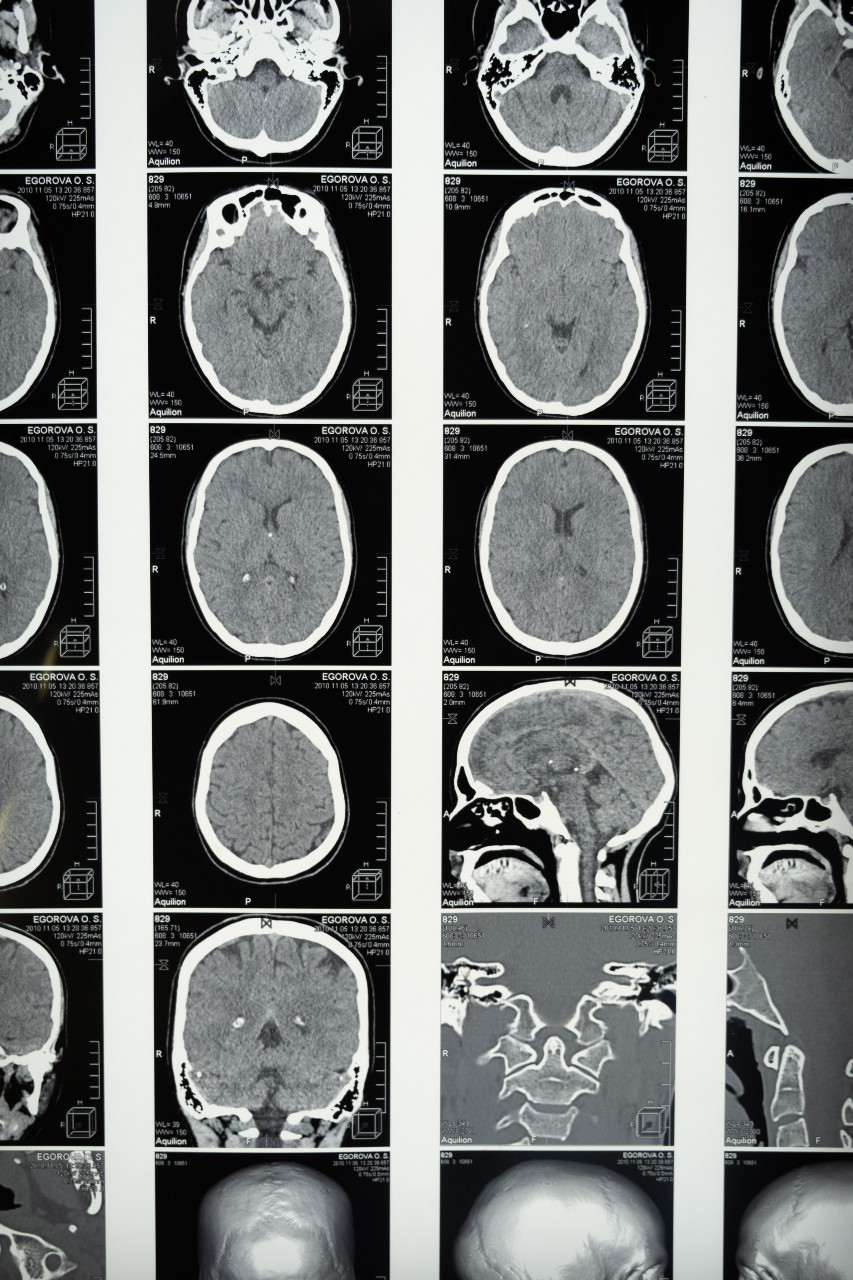
Traumatic Head Injury part 4
It is important to reduce any possibility of secondary brain injury occurring following a TBI:
- Secondary brain injury can be described as injury following the primary event, such as resulting from hypoxia, hypercarbia, or hypoperfusion. An example of this can be impact brain apnoea, where following a TBI a patient may have a prolonged period of apnoea, eventually leading to cardiac arrest. If the patients’ hypoxic needs can be managed by the clinician, this will avert secondary brain injury. Managing airway, breathing, and oxygenation of a patient can help halt hypoxia from being a factor of secondary brain injury.
- Carbon dioxide levels are also an important consideration due to its’ effects on the body. If hypercapnic, this causes vasodilation of cerebral blood vessels, increasing intercranial volumes and ICP. If hypocapnic, this causes vasoconstriction within the cerebral vasculature, worsening cerebral hypoxia and oedema. Managing a normocapnic level of 35 – 45 mmHg through ventilation rate can help minimise the negative effects of hyper and hypocapnia.
- Hypoperfusion can cause further secondary brain injury through not adequately perfusing the brain. Fluid therapy can be used to manage blood pressure levels and assist with maintaining MAP and CPP. If a patient has a TBI with other blunt trauma or penetrating limb trauma, then a systolic of 90 mmHg can be maintained. A targeted systolic of 60 mmHg or central carotid pulse should be aimed for in penetrating torso trauma, and a systolic of 110 mmHg in isolated head injury. A traumatic event causing a major head injury is likely to have caused trauma to other parts of the body. The neck and c-spine may have suffered significant damage and may also be compromised. Consideration for c-spine control will have to be taken when looking at moving the patient.
- On reviewing c-spine control, there is growing discussion on whether cervical collars should be used in instances of head injury with the risk of TBI. The reason for this is due to the risk of potential increased ICP generated through the collar putting pressure on the neck and venous return, causing further chance of swelling and ICP. A decision will need to be made if a collar is to be used, loosened, or removed if suspecting TBI. On reviewing this, different services have different guidelines, and it is important to review your own guidelines and follow what procedures and practices are present in your own ambulance service.
- Tranexamic Acid (TXA) is an important consideration when dealing with major head injury due to its’ potential to reduce further intercranial bleeding and death. Following your local drug guidelines, TXA may be administered to major head injury patients.
- TBI patient may present with significant agitation and irritability, making it difficult to manage. Through the patients’ own actions, they may also inadvertently further harm themselves. Early request for specialist support, such as Critical Care support for Midazolam or Rapid Sequence Intubation (RSI) may help you and the patient in their overall care.
Treatment:
When dealing with a major head injury and suspecting TBI, working through the primary survey will assist in managing a difficult scenario. Remembering the considerations for TBI will also help with the overall outcome of the patient.
Working through the primary survey:
Catastrophic Haemorrhage:
Stemming any catastrophic haemorrhage is vital to maintain any homeostatic stability of the patient. Hypovolaemia will further secondary brain injury and worsen the patients’ outcome.
Airway with C-spine consideration:
Maintaining a patent airway through a stepwise approach will help stem hypoxic secondary brain damage. Remember there may be a likely C-spine injury and the use of a cervical collar may have to be omitted or altered to stop further increase in ICP.
Breathing:
Management of breathing will further assist in reducing hypoxic damage. Use oxygen therapy where there is major trauma. Ventilation and recording of capnography can be used to maintain normocapnic levels and reduce the risk of further secondary brain injury through hyper or hypocapnia.
Circulation:
Review further haemorrhage control where there may be internal concerns. Apply pelvic binder, TXA for major head injury and bleeding following your ambulance service guidelines. Look at fluid administration to maintain systolic blood pressures to keep CPP. A targeted systolic of 60 mmHg or central carotid pulse should be aimed for in penetrating torso trauma, a systolic of 90 mmHg or peripheral radial pulse in blunt trauma or penetrating limb trauma, and a systolic of 110 mmHg in isolated head injury.
Disability:
Maintain patients’ temperature and cover to avoid heat loss, review blood glucose levels and treat any unusual findings, review GCS level, review any seizure activity and treat if required.
Evacuate:
Consider early evacuation of the patient to further care, request specialist support if needed and available. Consider a 30⁰ head up position when transporting the patient as this can reduce the effects of increased ICP. This may be difficult though within the pre-hospital setting and where a patient is immobilised. Use clinical judgement and consideration for the scenario and setting in front of you.
